
Using Deep Learning to Facilitate Scientific Image Analysis
March 16, 2018
Posted by Samuel Yang, Research Scientist, Google Accelerated Science Team
Many scientific imaging applications, especially microscopy, can produce terabytes of data per day. These applications can benefit from recent advances in computer vision and deep learning. In our work with biologists on robotic microscopy applications (e.g., to distinguish cellular phenotypes) we've learned that assembling high quality image datasets that separate signal from noise is a difficult but important task. We've also learned that there are many scientists who may not write code, but who are still excited to utilize deep learning in their image analysis work. A particular challenge we can help address involves dealing with out-of-focus images. Even with the autofocus systems on state-of-the-art microscopes, poor configuration or hardware incompatibility may result in image quality issues. Having an automated way to rate focus quality can enable the detection, troubleshooting and removal of such images.
Deep Learning to the Rescue
In “Assessing Microscope Image Focus Quality with Deep Learning”, we trained a deep neural network to rate the focus quality of microscopy images with higher accuracy than previous methods. We also integrated the pre-trained TensorFlow model with plugins in Fiji (ImageJ) and CellProfiler, two leading open source scientific image analysis tools that can be used with either a graphical user interface or invoked via scripts.
 |
| A pre-trained TensorFlow model rates focus quality for a montage of microscope image patches of cells in Fiji (ImageJ). Hue and lightness of the borders denote predicted focus quality and prediction uncertainty, respectively. |
What about Images without Objects?
An interesting challenge we overcame was that there are often "blank" image patches with no objects, a scenario where no notion of focus quality exists. Instead of explicitly labeling these "blank" patches and teaching our model to recognize them as a separate category, we configured our model to predict a probability distribution across defocus levels, allowing it to learn to express uncertainty (dim borders in the figure) for these empty patches (e.g. predict equal probability in/out-of-focus).
What's Next?
Deep learning-based approaches for scientific image analysis will improve accuracy, reduce manual parameter tuning and may reveal new insights. Clearly, the sharing and availability of datasets and models, and implementation into tools that are proven to be useful within respective communities, will be important for widespread adoption.
Acknowledgements
We thank Claire McQuin, Allen Goodman, Anne Carpenter of the Broad Institute and Kevin Eliceiri of the University of Wisconsin at Madison for assistance with CellProfiler and Fiji integration, respectively.
Other posts of interest
-
April 19, 2024
Improving Gboard language models via private federated analytics- Algorithms & Theory ·
- Distributed Systems & Parallel Computing ·
- Product ·
- Security, Privacy and Abuse Prevention
-
April 12, 2024
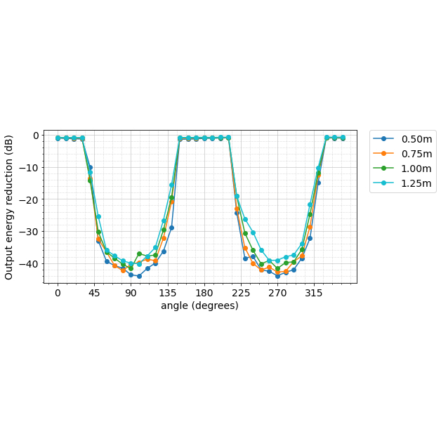
Contrastive neural audio separation- Machine Intelligence ·
- Sound & Accoustics
-
April 11, 2024
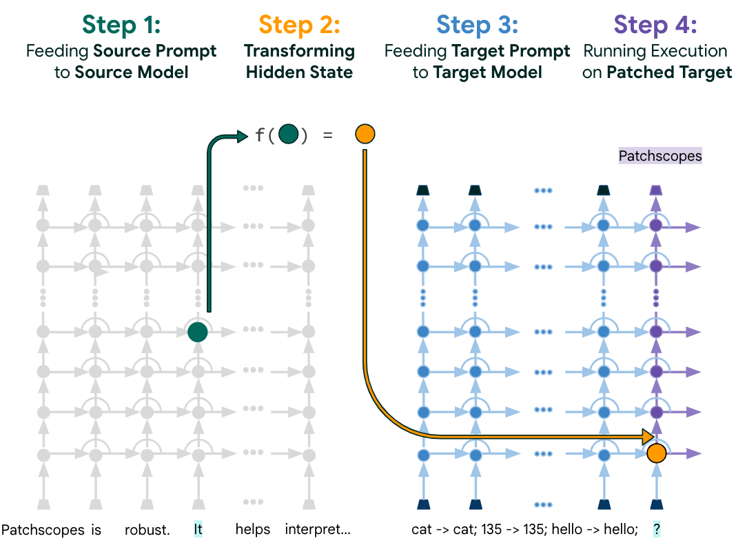
Patchscopes: A unifying framework for inspecting hidden representations of language models- Machine Intelligence ·
- Natural Language Processing ·
- Responsible AI